
Mammography
What is Mammography and what does it do?
Mammography is a low-dose radiological examination allowing breast problems to be detected, such as benign abnormalities or the development of tumors. Examination involves an X-Ray machine that has been specifically designed to display the structure of the breast.
Jackson Hospital uses a full- field digital mammography system which uses a digital detector. This is the latest generation of mammography equipment. The system generates digital 2D or 3D images that can be used for screening and diagnostic imaging. It offers new capabilities of processing images, such as, zoom, contrast adjustment, and computer- aided detector (CAD). Digital signals are sent to a computer-aided detector (CAD) reader, also known as a second reader. The computer pre-reads the mammograms, identifying areas of suspicion or areas needing additional workup.
Mammographic studies include:
Mammogram (Diagnostic)
What is a Diagnostic Mammogram and what does it do?
Diagnostic mammography is used to evaluate a patient with abnormal clinical findings—such as a breast lump or lumps—that have been found by the woman or her doctor. Diagnostic mammography may also be done after an abnormal screening mammogram in order to evaluate the area of concern on the screening exam.
Who performs the test?
The examination is performed by a licensed Radiologic Technologist.
Where does it take place?
Jackson Hospital Outpatient Center, Hudnall Building, Room 110, located adjacent to the Hospital
How long does it take?
Average person 15-30 minutes
What you can do to make it a success?
- Please bring physician orders to your appointment.
- Patients who are in a wheelchair, please ensure your wheelchair arms and legs can be removed. If not, be prepared to be moved to a different chair.
- Inform technologist prior to exam if there is a possibility of pregnancy.
What to do before your exam?
On the day of your appointment, do not use deodorant, lotion, cream, or powder on your underarms or breasts, as they could interfere with a clear image. Also, wear clothing that will allow you to undress from the waist up easily.
If you still have a menstrual cycle, please schedule you mammogram for one week after your period ends.
If you have had a mammogram at another facility, have previous mammograms available for the radiologist at your current exam.
You will be asked to fill out a questionnaire concerning breast health history.
What happens during your exam?
Your breast will be placed on a special platform and compressed with a paddle (often made of clear Plexiglas or other plastic). The technologist will gradually compress your breast.
Breast compression is necessary in order to:
- Even out the breast thickness so that all of the tissue can be visualized.
- Spread out the tissue so that small abnormalities are less likely to be obscured by overlying breast tissue.
- Allow the use of a lower x-ray dose since a thinner amount of breast tissue is being imaged.
- Hold the breast still in order to minimize blurring of the image caused by motion.
- Reduce x-ray scatter to increase sharpness of picture.
- You will be asked to change positions between images. The routine views are a top-to-bottom view and an angled side view. The process will be repeated for the other breast.
- You must hold very still and may be asked to keep from breathing for a few seconds while the x-ray picture is taken to reduce the possibility of a blurred image. The technologist will walk behind a wall or into the next room to activate the x-ray machine.
- Ultrasound and/or further examination by mammography may be warranted.
What to do after your exam?
A radiologist will then examine the images and interpret them. He will communicate the results and forward a report to your doctor. The patient will receive notification through a letter from the Mammogram facility. If the previous mammograms are not provided at the current exam, there will be a delay in notifying the ordering physician as well as the patient of the results as we locate and compare previous and current mammograms.
Contact Information:
Hospital (main operator): 850-526-2200
Mammography Department at OP Center: 850-718-2585 or 850-718-
2595
Imaging Services Department (at hospital): 850-718-2580
Mammogram (Screening)
What is a screening mammogram and what does it do?
A screening mammogram refers to the medical screening of asymptomatic (having no symptoms), apparently healthy women for breast cancer in an attempt to achieve an earlier diagnosis. The belief is that early detection will improve outcomes
Who performs the test?
The examination is performed by a licensed Radiologic Technologist certified in Mammography.
Where does it take place?
Jackson Hospital Outpatient Center Hudnall Building, Room 110, located adjacent to the Hospital
How long does it take?
Average person 10-15 minutes
What you can do to make it a success?
- Please bring physician orders to your appointment.
- Patients who are in a wheelchair, please ensure your wheelchair arms and legs can be removed. If not, be prepared to be moved to a different chair.
- Inform technologist prior to exam if there is a possibility of pregnancy.
What to do before your exam?
On the day of your appointment, do not use deodorant, lotion, cream, or powder on your underarms or breasts, as they could interfere with a clear image. Also, wear clothing that will allow you to undress from the waist up easily.
If you still have a menstrual cycle, please schedule you mammogram for one week after your period ends.
If you have had a mammogram at another facility, have previous mammograms available for the radiologist at your current exam.
You will be asked to fill out a questionnaire concerning breast health history.
What happens during your exam?
Your breast will be placed on a special platform and compressed with a paddle (often made of clear Plexiglas or other plastic). The technologist will gradually compress your breast.
Breast compression is necessary in order to:
- Even out the breast thickness so that all of the tissue can be visualized.
- Spread out the tissue so that small abnormalities are less likely to be obscured by overlying breast tissue.
- Allow the use of a lower x-ray dose since a thinner amount of breast tissue is being imaged.
- Hold the breast still in order to minimize blurring of the image caused by motion.
- Reduce x-ray scatter to increase sharpness of picture.
- You will be asked to change positions between images. The routine views are a top-to-bottom view and an angled side view. The process will be repeated for the other breast.
- You must hold very still and may be asked to keep from breathing for a few seconds while the x-ray picture is taken to reduce the possibility of a blurred image. The technologist will walk behind a wall or into the next room to activate the x-ray machine.
- Ultrasound and/or further examination by mammography may be warranted.
What to do after your exam?
A radiologist will then examine the images and interpret them. He will communicate the results and forward a report to your doctor. The patient will receive notification through a letter from the Mammogram facility. If the previous mammograms are not provided at the current exam, there will be a delay in notifying the ordering physician, as well as, the patient of the results as we locate and compare previous and current mammograms.
Contact Information:
Hospital (main operator): 850-526-2200
Mammography Department at OP Center: 850-718-2585 or 850-718-2595
Imaging Services Department (at hospital): 850-718-2580
Mammogram (Implants)
What is a Mammogram of Implants and what does it do?
A woman with implants should follow the same breast-care guidelines as a woman without implants. If you have implants, then the mammograms must be done in a special way in order to see the breast tissue effectively, so you have to tell the technician that you have implants. Mammograms take more time to perform when there are implants, because extra images are needed, the technique is different and extra care must be taken when compressing the breast to reduce the risk of implant rupture.
Special views will be done when implants are present. These techniques are called breast implant displacement views, Eklund displacement views, or Eklund views, named for the radiologist who developed the techniques. These special implant displacement views are done in addition to those views done during routine mammograms.
The displacement procedure involves pushing the implant back and pulling the breast tissue into view. Several factors, which may affect the success of this special technique in imaging the breast tissue in women with breast implants, include the location of the implant, the hardness of the capsular contracture and the amount of the breast tissue compared to the implant size.
Who performs the test?
The examination is performed by a licensed Radiologic Technologist.
Where does it take place?
Jackson Hospital Outpatient Center Hudnall Building, Room 110, located adjacent to the Hospital
How long does it take?
Average person 15-30 minutes
What you can do to make it a success?
- Please bring physician orders to your appointment.
- Patients who are in a wheelchair, please ensure your wheelchair arms and legs can be removed. If not, be prepared to be moved to a different chair.
- Inform technologist prior to exam if there is a possibility of pregnancy.
What to do before your exam?
On the day of your appointment, do not use deodorant, lotion, cream, or powder on your underarms or breasts, as they could interfere with a clear image. Also, wear clothing that will allow you to undress from the waist up easily.
If you still have a menstrual cycle, please schedule you mammogram for one week after your period ends.
If you have had a mammogram at another facility, have previous mammograms available for the radiologist at your current exam.
You will be asked to fill out a questionnaire concerning breast health history.
What happens during your exam?
Your breast will be placed on a special platform and compressed with a paddle (often made of clear Plexiglas or other plastic). The technologist will gradually compress your breast.
Breast compression is necessary in order to:
- Even out the breast thickness so that all of the tissue can be visualized.
- Spread out the tissue so that small abnormalities are less likely to be obscured by overlying breast tissue.
- Allow the use of a lower x-ray dose since a thinner amount of breast tissue is being imaged.
- Hold the breast still in order to minimize blurring of the image caused by motion.
- Reduce x-ray scatter to increase sharpness of picture.
- You will be asked to change positions between images. The routine views are a top-to-bottom view and an angled side view. The process will be repeated for the other breast.
- You must hold very still and may be asked to keep from breathing for a few seconds while the x-ray picture is taken to reduce the possibility of a blurred image. The technologist will walk behind a wall or into the next room to activate the x-ray machine.
- Ultrasound and/or further examination by mammography may be warranted.
What to do after your exam?
A radiologist will then examine the images and interpret them. He will communicate the results and forward a report to your doctor. The patient will receive notification through a letter from the Mammogram facility. If the previous mammograms are not provided at the current exam, there will be a delay in notifying the ordering physician, as well as, the patient of the results as we locate and compare previous and current mammograms.
Contact Information:
Hospital (main operator): 850-526-2200
Mammography Department at OP Center: 850-718-2585 or 850-718-2595
Imaging Services Department (at hospital): 850-718-2580
Needle Localization
What is a Needle Localization and what does it do?
Needle Localization is a pre-operative procedure used to establish the location of a lesion that is to be removed by a Surgeon while the patient is under anesthesia. A wire marker is left in place in the breast by the Radiologist.
Who performs the test?
The examination is performed by a licensed Radiologic Technologist and a Radiologist.
Where does it take place?
(OP Center) Hudnall Building Room 110, located adjacent to the Hospital
How long does it take?
Average person 30 minutes to 1 hour
What to do before your exam?
The Needle Localizations/Biopsy will be scheduled through Outpatient Surgery and your Surgeon. They will advise you of pre-op instructions. Loose clothing recommended.
What happens during your exam?
The technologist takes X-rays of your breast to help localize the abnormality during surgery.
The Radiologist will administer a local anesthetic to numb the site before he inserts a needle fitted with a wire and marker in the area located.
A further X-ray is then taken to check the position of the marker.
The Radiologist withdraws the needle, leaving the marker at the end of the wire which is protected by a dressing. The marker remains in place until the surgeon intervenes.
What to do after your exam?
Reduce activity and follow post-op instructions from Outpatient Surgery and your Surgeon.
Contact Information:
Hospital (main operator): 850-526-2200
Mammography Department at OP Center: 850-718-2585 or 850-718-2595
Imaging Services Department (at hospital): 850-718-2580
Ultrasound Breast Biopsy
What is an Ultrasound Guided Breast Biopsy and what does it do?
Lumps or abnormalities in the breast are often detected by physical examination, mammography, or other imaging studies. However, it is not always possible to tell from these imaging tests whether a growth is benign or cancerous.
A breast biopsy is performed to remove some cells—either surgically or through a less invasive procedure involving a hollow needle—from a suspicious area in the breast and examine them under a microscope to determine a diagnosis. Image-guided needle biopsy is not designed to remove the entire lesion, but most of a very small lesion may be removed in the process of biopsy.
In ultrasound-guided breast biopsy, ultrasound imaging is used to help guide the doctor’s instruments to the site of the abnormal growth.
An ultrasound-guided breast biopsy can be performed when a breast ultrasound shows an abnormality such as:
- a suspicious solid mass
- a distortion in the structure of the breast tissue
- an area of abnormal tissue change
One of four instruments will be used:
- A fine needle attached to a syringe, smaller than needles typically used to draw blood.
- A core needle, also called an automatic, spring-loaded needle, which consists of an inner needle connected to a trough, or shallow receptacle, covered by a sheath and attached to a spring-loaded mechanism.
- A vacuum-assisted device (VAD), a vacuum-powered instrument that uses pressure to pull tissue into the needle.
- A thin guide wire, which is used for a surgical biopsy.
Who performs the test?
The biopsy itself is performed by a physician with the assistance of an ultrasonographer specifically trained or certified in Ultrasound imaging.
Where does it take place?
At Jackson Hospital in the Imaging Services Department.
How long does it take?
This exam generally takes about 45 minutes to complete.
What you can do to make it a success?
Prior to a needle biopsy, you should report to your doctor all medications that you are taking, including herbal supplements, and if you have any allergies, especially to anesthesia. Your physician will advise you to stop taking aspirin or a blood thinner three days before your procedure.
Also, inform your doctor about recent illnesses or other medical conditions.You may want to have a relative or friend accompany you and drive you home afterward.
What to do before your exam?
No special preparation is needed for this study.
What happens during your exam?
Breast biopsies are usually done on an outpatient basis.
You will be positioned lying face up on the examination table or turned slightly to the side.
A local anesthetic will be injected into the breast to numb it.
Pressing the transducer to the breast, the sonographer or doctor will locate the lesion.
A very small nick is made in the skin at the site where the biopsy needle is to be inserted.
The doctor, monitoring the lesion site with the ultrasound probe, will insert the needle and advance it directly into the mass.
Tissue samples are then removed using one of three methods:
- In a fine needle aspiration, a fine gauge needle and a syringe withdraw fluid or clusters of cells.
- In a core needle biopsy, the automated mechanism is activated, moving the needle forward and filling the needle trough, or shallow receptacle, with ‘cores’ of breast tissue. The outer sheath instantly moves forward to cut the tissue and keep it in the trough. This process is repeated three to six times.
- With a vacuum-assisted device (VAD), vacuum pressure is used to pull tissue from the breast through the needle into the sampling chamber. Without withdrawing and reinserting the needle, it rotates positions and collects additional samples. Typically, eight to 10 samples of tissue are collected from around the lesion.
After this sampling, the needle will be removed.
If a surgical biopsy is being performed, a wire is inserted into the suspicious area as a guide for the surgeon.
A small marker may be placed at the site so that it can be located in the future if necessary.
Once the biopsy is complete, pressure will be applied to stop any bleeding and the opening in the skin is covered with a dressing. No sutures are needed.
A mammogram may be performed to confirm that the marker is in the proper position.
This procedure is usually completed within an hour.
What to do after your exam?
You will be given detailed discharge instructions to take home with you. These will have your after care instructions, what to watch for and contact information.
Contact Information:
Ultrasound Department (at main hospital): 850-718-2582
Ultrasound Department (at OP Center): 850-526-6702
Imaging Services Department: 850-718-2580
Hospital (main operator): 850-526-2200
Breast Self Exam (BSE)
What is (BSE) breast self-examination?
Breast Self examination is a screening method used in an attempt to detect early breast cancer. The method involves the woman herself looking at and feeling each breast for possible lumps, distortions or swelling.
Carried out regularly, BSE will allow you to become familiar with the normal appearance of your breasts, and will make your examinations more effective.
3 Steps that could save your life
In the shower
Examine your breasts in the shower or the bath. Hands move more easily over wet skin. With your fingers stretched, move your hand over the whole surface of your breasts. Look out for any unusual lumps, nodules or thickening.
In front of the mirror
Examine your breasts with your arms alongside your body. Then raise your arms above your head. Look for any changes in the shape of your breasts, for swellings or depressions in the skin, as well as for any changes to the nipples. Then place your hands on your hips, and push firmly downwards to stretch your pectoral muscles. There are very few women who have two identical breasts.
Lying down
To examine your right breast, place a pillow or a folded towel underneath your right shoulder. Place your right hand behind your head in order to improve the distribution of the breast tissue on your chest. Using your left hand, palpate the breast by making concentric circles, as if you were following the outline of a clock face. First of all follow the outermost circle. Start palpating the imaginary clock dial at the twelve o’clock location. Slide your fingers to one o’clock, and so on.
The presence of firmer tissue in the lower part of the breast is quite normal. To start following a new concentric circle, move your fingers about 2cm towards your nipple, and in this way examine the whole of your breast, including the nipple. In order to palpate the entire breast, you should have to make about five circles.
Repeat this procedure for the left breast by placing a pillow under your left shoulder and your left hand behind your head. Finally press the nipple of both breasts gently between your thumb and index finger. If there is any discharge, you should see your doctor immediately.
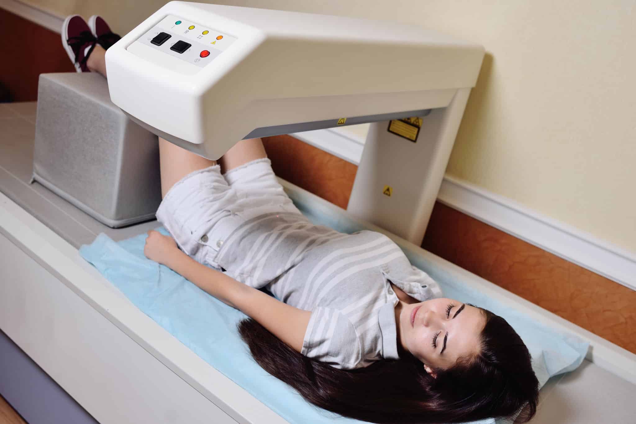
DEXA Scan (HIP/SPINE/FOREARM)
Bone density scanning, also called dual-energy x-ray absorptiometry (DEXA) or bone densitometry, is an enhanced form of x-ray technology that is used to measure bone loss. The device has a large, flat table and an “arm” suspended overhead.
DEXA is recognized as the standard for measuring bone mineral density (BMD). DEXA is most often performed on the lower spine and hips, unless a prosthesis is present in the hip or lower spine. When prosthesis is present in either area a scan of the forearm will be performed and analyzed.
DEXA studies include:
DEXA (hip/spine/forearm)
What is a DEXA scan and what does it do?
DEXA stands for ‘Dual Energy X-ray Absorptiometry’. It is the most commonly used test for measuring bone mineral density. It is one of the most accurate ways to diagnosis Osteopenia or Osteoporosis.
A DEXA scan detects weak or brittle bones before you have a fracture. The score helps to predict your chance of fracture in the future, and perhaps the need for osteoporosis medication. The DEXA scan, when compared to previous DEXA scan results, indicates whether your bone density is improving, worsening, or staying the same. It helps determine if your osteoporosis medication is working. After a fracture occurs, a DEXA scan can assess if it was likely due to osteoporosis.
Who performs the test?
The examination is performed by a licensed Radiologic Technologist.
Where does it take place?
Outpatient Center Hudnall Building, Room 110, located adjacent to the hospital
How long does it take?
Average person 10-15 minutes
What you can do to make it a success?
- Please bring physician orders to your appointment.
DEXA exam table weight limit is 300 lbs
Inform technologist prior to exam if there is a possibility of pregnancy.
What to do before your exam?
On the day of the exam you may eat normally. You should not take calcium supplements for at least 24 hours before your exam. You should wear loose, comfortable clothing, avoiding garments that have zippers, belts or buttons made of metal. You will be asked to remove objects such as keys or wallets that would be in the area being scanned. You may be asked to remove some or all of your clothes and to wear a gown during the exam.
You will have to wait 3 to 7 days before undergoing a DEXA test, if you recently had a barium examination or have been injected with a contrast material for a computed tomography scan (CT) or radioisotope scan as these would interfere with your results.
What happens during your exam?
In the DEXA examination, which measures bone density in the hip and spine, the patient lies on a padded table. If you are unable to lie on the table or have metallic implants in your hips and/or spine that may interfere with your results, you can sit in a chair with your forearm on the scanning table for a DEXA scan of the forearm.
An imaging device, or detector, is positioned above which slowly passes over the area, generating images on a computer monitor.
You must hold very still while the x-ray picture is taken to reduce the possibility of a blurred image.
What to do after your exam?
A Radiologist will analyze the images and send a signed report to your physician.
Your test results will be in the form of two scores:
- T score — This number shows the amount of bone you have compared with a young adult of the same gender with peak bone mass. A score above -1 is considered normal. A score between -1 and -2.5 is classified as osteopenia (low bone mass). A score below -2.5 is defined as osteoporosis. The T score is used to estimate your risk of developing a fracture.
- Z score — This number reflects the amount of bone you have compared with other people in your age group and of the same size and gender. If this score is unusually high or low, it may indicate a need for further medical tests.
Contact Information:
Hospital (main operator): 850-526-2200
Mammography Department at OP Center: 850-718-2585 or 850-718-2595
Imaging Services Department (at hospital): 850-718-2580
DEXA (body composition)
What is a DEXA Body Composition scan and what does it do?
DEXA stands for (Dual Energy X-ray Absorptiometry) which is used to assess your bone density and well as your body composition. DEXA uses an x-ray technique to look at the density of the body and can then estimate the amount of lean muscle mass and fat tissue.
Importance of Your Body Fat Percentage
A person can have a lot of muscle, but be considered “over-weight” by many height/weight charts. The opposite can also be true – a person can have a lot of fat and little muscle and be “over-fat” but not overweight.
Too little body fat is linked to problems with normal, healthy functioning in both men and women. It can also lead to problems with reproduction in women.
Too much body fat, especially when located around the abdomen, increases the risk of many diseases, including the following:
- Type II diabetes
- High blood pressure
- Stroke
- Heart disease
- Certain cancers
- For many athletes, performance is improved with optimal body composition.
By knowing your body fat percentage, and the range it falls in, you can take measures to improve your health and reduce your risk for various diseases.
Who performs the test?
The examination is performed by a licensed Radiologic Technologist.
Where does it take place?
Outpatient Center Hudnall Building, Room 110, located adjacent to the hospital.
How long does it take?
Average scan takes about 20 minutes.
What you can do to make it a success?
DEXA exam table weight limit is 300 lbs and height limit is 6’. If you weigh over 300 pounds or are taller than 6 foot, you cannot have a Body Composition Scan.
Inform technologist prior to exam if there is a possibility of pregnancy.
You will have to wait 3 to 7 days before undergoing a DEXA study if you recently had a barium examination or have been injected with a contrast material for a computed tomography scan (CT) or radioisotope scan (Nuclear Medicine) as these would interfere with your results.
What should I do before the exam?
On the day of the exam you may eat normally. You should wear loose, comfortable clothing, avoiding garments that have zippers, belts or buttons made of metal. You will be asked to remove objects such as keys or wallets that would be in the area being scanned. You may be asked to remove some or all of your clothes and to wear a gown during the exam.
What happens during the exam?
You will be asked to lie down on a padded table. An imaging device, or detector, is positioned above you and slowly passes over your body several times, generating images on a computer monitor. You must hold very still while the x-ray picture is taken to reduce the possibility of a blurred image.
What should I do after the exam?
You may resume your normal activity after your exam. Your body composition results will be given to you to keep.
WE DO NOT SEND A COPY TO YOUR PHYSICIAN.
Contact Information:
To schedule: (850) 718-2552
DEXA Department at OP Center: (850) 718-2585 or (850) 718-2595
Imaging Services Department (at hospital): (850) 718-2580
Where are Mammo/Dexa Services located?
Mammo and Dexa are located in the Hudnall Medical Building, adjacent to Jackson Hospital, in the Outpatient Center, Room 110.