X-Ray Services
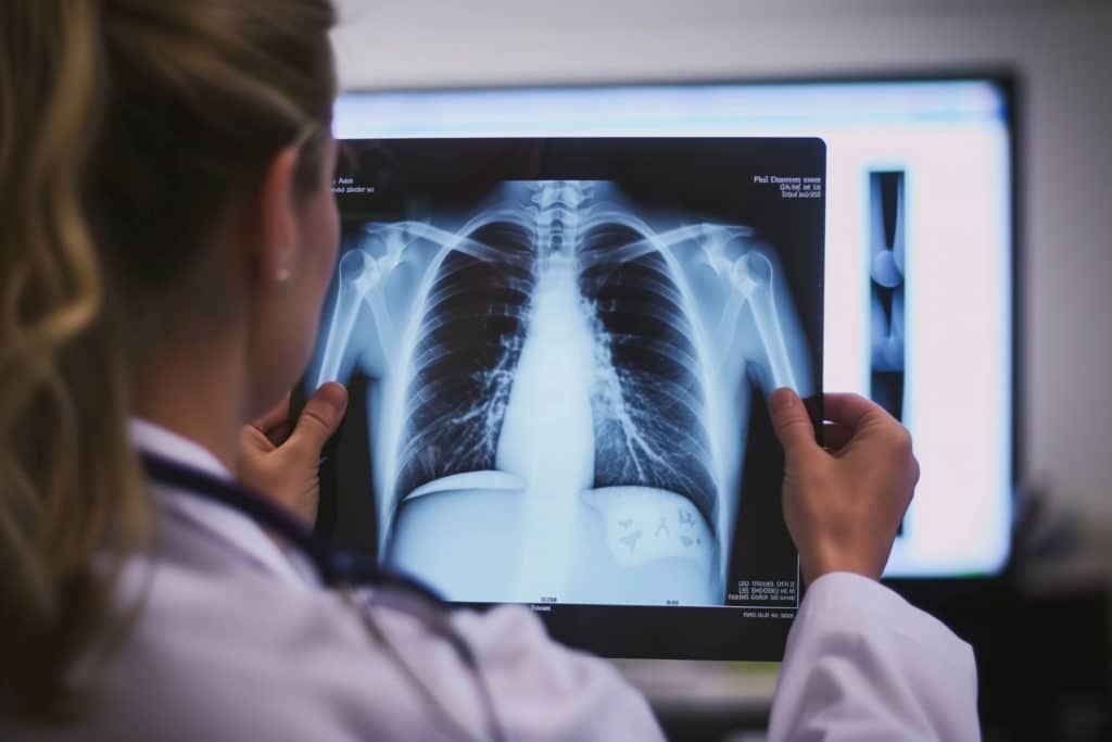
An X-Ray is a painless imaging test that uses small amounts of radiation to produce 2-D images of the structures inside your body. X-Ray images help physicians view structures within the body such as bones, which show up as white, and fat and muscle, which show up as varying shades of gray. These X-Ray images help physicians diagnose certain diseases and show bone fractures.
General X-ray studies include images of the:
- Chest
- Abdomen
- Pelvis
- Sinuses
- Skull
- Spine (Cervical/Lumbar/Thoracic)
- Upper Extremities (Shoulder, Humerus, Elbow, Forearm, Wrist, Hand, Fingers)
- Lower Extremities (Hip, Femur, Knee, Tibia/Fibula, Ankle, Foot, Toes)
Most general x-ray procedures require no special preparation. Once you arrive, you may be asked to change into a gown before your examination. You will also be asked to remove jewelry, eyeglasses and any metal objects that could show up on the images and overlap important findings.
Women should always inform their doctor or X-ray technologist if there is any possibility that they are pregnant.
Where is the X-Ray Department Located?
At Jackson Hospital in the Imaging Services Department
Imaging Services:
Imaging studies include but are not limited to the following items listed:
Barium Swallow (BaS)
What is a Barium Swallow and what does it do?
- A barium esophagram, or barium swallow, may be ordered for patients with difficult or painful swallowing, coughing, choking, a sensation of something stuck in the throat, or chest pain. The test is performed when a patient drinks the barium and X-ray images are taken. These problems can be detected with a barium swallow:
- Narrowing or irritation of the esophagus (the muscular tube between the back of the throat and the stomach)
- Disorders of swallowing
- Hiatal hernia (an internal defect that causes the stomach to slide partially into the chest)
- Abnormally enlarged veins in the esophagus that cause bleeding
- Ulcers
- Tumors
- Polyps (growths that are usually not cancerous, but could be precancerous)
Who performs the test?
The examination is performed by a doctor specially trained in Radiology (Radiologist) and a licensed Radiologic Technologist RT (R).
Where does it take place?
At Jackson Hospital in the Imaging Services Department
How long does it take?
Average person 15-30 minutes
What you can do to make it a success?
- Wear comfortable, easy to remove clothing.
- Patients will be required to put a gown on for all Fluoroscopy x-ray exams.
- Follow all preparation instructions given to you by your physician’s office. If you have any questions, please call us for clarification. We want your exam to be as successful as possible.
What to do before your exam?
- Take nothing by mouth 8 hours prior to your exam. You may take your medications with minimal amount water.
- If you are a woman of childbearing age and there is a chance you may be pregnant, please consult your physician before scheduling this exam.
What happens during your exam?
- You will be asked to dress in a patient gown.
- You will be given a small cup of carbonated water and a cup of liquid barium to drink while the radiologist observes under fluoroscopy and takes images of your esophagus.
- You will be positioned for your exam based on the area of the body to be x-rayed. This could be standing, sitting or lying down in various positions on the exam table.
- Most exams require multiple views or positions of the body part for adequate evaluation.
What to do after your exam?
- You will be given discharge instructions requesting that a mild laxative be taken after the study.
- After the examination, your stool will be lightly colored from the barium for 24 to 72 hours. It is important to remove the barium from the large intestine. If the barium is not removed, it may harden and block the intestine. Drink 6 to 8 glasses (soda pop can size) of liquid after the test to help get rid of the barium. This will also help to keep you from being constipated or dehydrated.
Colonic Transit Study
What is a Colonic Transit Study and what does it do?
A Colonic Transit Study is used by your doctor to diagnose severe constipation or other colon disorders. You will be given a tablet (Jackson Hospital uses Sitzmarks® brand) to swallow. The tablet contains tiny plastic rings that will pass naturally through the digestive tract just as food does. An x-ray will be taken 5 days after the tablet was taken to determine the amount of rings left in your digestive tract. This will help your doctor determine what type of condition you may have and how to treat it.
Who performs the test?
The exam itself is performed by a Radiologic Technologist RT (R). These technologists are nationally registered with the A.R.R.T. (American Registry of Radiologic Technologists) and licensed through the state of Florida.
Where does it take place?
At Jackson Hospital in the Imaging Services Department
How long does it take?
You must come to the hospital to receive your tablet (we prefer you take the tablet at that time).
You will then be given a day and time (5 days after taking tablet) to return for your abdomen x-ray.
The x-ray itself takes about 15 minutes.
What you can do to make it a success?
Be sure to return on the day and time you were given.
Bring your orders with you when you come for your x-ray.
If you are pregnant, please let your physician know BEFORE you come to hospital for your tablet.
What to do before your exam?
It is recommended that you wear loose, comfortable clothing for the exam. You will need to remove any metallic objects that may be in the path of the x-ray beam (belts, zippers, piercings, etc). To reduce the risk of valuables being lost, it is recommended that they be removed prior to entering the exam room or simply left at home.
Do not use any laxatives until you have been informed that your test is complete. Laxatives include stimulants, bulk forming fibers, suppositories and saline enemas. You may take your prescribed medications.
There are no dietary restrictions for this test.
What happens during your exam?
You may have to change into a hospital gown for your study.
You will be asked to lie on an exam table for your abdominal x-ray. Once the x-ray is taken, you will be asked to wait while the doctor looks at your picture. If there are still several markers present in your digestive system, you may be asked to return in a couple of days for another x-ray (Day 7). If most of the markers have passed the exam is complete. If not, you may be instructed to come back in 3 days (Day 10) for another abdominal x-ray. Day 10 will be the last day.
What to do after your exam?
If you are not instructed to come back for additional x-rays, you will be released to go home. The radiologist will review your image(s) and a final report will go to your ordering physician in 24-48 hours.
Gastrointestinal Radiology
Gastroenterologists often order radiographic tests to help diagnose diseases of the gastrointestinal tract. Common complaints that may lead to such testing include abdominal pain, nausea, vomiting, heartburn, diarrhea, constipation, blood in the stool, bloating, weight loss, and abnormal laboratory tests.
Fluoroscopy is an imaging technique that uses X-rays to obtain real-time moving images of the internal structures of a patient through the use of a fluoroscope allowing the images to be recorded and played on a monitor.
Barium Sulfate is commonly used to allow for better visualization of gastrointestinal organs. Barium sulfate is a harmless chalky, water-insoluble compound that does not permit x-rays to pass through it. Taken before or during an examination, it causes the intestinal tract to stand out in silhouette when viewed through a fluoroscope or seen on an x-ray film. It is important to evacuate the barium completely following the study; a mild laxative is usually prescribed for this purpose.
Gastrointestinal studies include:
- Barium Swallow (BaS)
- Barium Swallow (BaS) Modified w/ Speech Therapist
- Upper GI (Gastrointestinal) Series
- Lower GI (Gastrointestinal) Series
- Small Bowel Series (SBS)
- Colonic Transit Study
Upper GI (Gastrointestinal) Study
What is an Upper GI and what does it do?
An upper gastrointestinal series is a barium study evaluating the esophagus, stomach, and first part of the small intestine. This test is ordered to search for causes of nausea, vomiting, abdominal pain, or weight loss, to name a few. It is performed much the same way as the barium esophagram, except additional time is required to take pictures as the barium travels further in the intestinal tract.
Who performs the test?
The examination is performed by a doctor specially trained in Radiology (Radiologist) and a licensed Radiologic Technologist RT (R).
Where does it take place?
At Jackson Hospital in the Imaging Services Department
How long does it take?
Average person 15-30 minutes
What you can do to make it a success?
- Wear comfortable, easy to remove clothing.
- Follow all preparation instructions given to you by your physician’s office. If you have any questions, please call us for clarification. We want your exam to be as successful as possible.
What to do before your exam?
- Take nothing by mouth 8 hours prior to your exam. You may take your medications with minimal amount water.
- If you are a woman of childbearing age and there is a chance you may be pregnant, please consult your physician before scheduling this exam.
What happens during your exam?
- You will be asked to dress in a patient gown.
- You will be given a small cup of carbonated water and a cup of liquid barium to drink while the radiologist observes under fluoroscopy and takes images of your esophagus, stomach and duodenum.
- You will be positioned for your exam based on the area of the body to be x-rayed. This could be standing, sitting or lying down in various positions on the exam table.
- Most exams require multiple views or positions of the body part for adequate evaluation.
What to do after your exam?
- You will be given discharge instructions requesting that a mild laxative be taken after the study.
- After the examination, your stool will be lightly colored from the barium for 24 to 72 hours. It is important to remove the barium from the large intestine. If the barium is not removed, it may harden and block the intestine. Drink 6 to 8 glasses (soda pop can size) of liquid after the test to help get rid of the barium. This will also help to keep you from being constipated or dehydrated.
- Colostomy (ko-loss-tuh-mee): If you have a colostomy, irrigate it after the last x-ray is taken and again the following morning.
Small Bowel Series (SBS)
What is a Small Bowel Series and what does it do?
A small bowel follow through (SBFT) or Small bowel series (SBS) is a fluoroscopic barium study of the small intestine. This test is usually ordered in conjunction with the Upper GI Series (UGI). The patient drinks a contrast medium containing barium sulfate. This contrast medium appears white on x-rays, and shows the outline of the internal lining of the bowel. X-ray images are taken as the contrast moves through the intestine, commonly at 0 minutes, 30 minutes, 60 minutes and 90 minutes. This enables the radiologist to assess the bowel as it becomes visible. The test is completed when the Barium is visualized in the terminal ileum and Cecum, which marks the beginning of the large bowel. This is one of the most common places for pathology of the bowel to be found; therefore imaging of this structure is crucial. The test length varies from patient to patient as bowel motility is highly variable.
Who performs the test?
The examination is performed by a licensed Radiologic Technologist RT (R).
Where does it take place?
At Jackson Hospital in the Imaging Services Department
How long does it take?
Average person takes 2-4 hours; maybe longer if there are motility problems.
What you can do to make it a success?
- Wear comfortable, easy to remove clothing.
- Follow all preparation instructions given to you by your physician’s office. If you have any questions, please call us for clarification. We want your exam to be as successful as possible.
What to do before your exam?
- Take nothing by mouth 8 hours prior to your exam. You may take your medications with minimal amount water.
- If you are a woman of childbearing age and there is a chance you may be pregnant, please consult your physician before scheduling this exam.
What happens during your exam?
- You will be asked to dress in a patient gown.
- You will be given liquid barium to drink.
- The technologist will take images of your abdomen at timed intervals to track the barium through your small bowel.
- Once the barium has reached the terminal ilieum and cecum, the exam is complete.
What to do after your exam?
- You will be given discharge instructions requesting that a mild laxative be taken after the study.
- After the examination, your stool will be lightly colored from the barium for 24 to 72 hours. It is important to remove the barium from the large intestine. If the barium is not removed, it may harden and block the intestine. Drink 6 to 8 glasses (soda pop can size) of liquid after the test to help get rid of the barium. This will also help to keep you from being constipated or dehydrated.
Lumbar Puncture
Lumbar Puncture (Spinal Tap) at Jackson Hospital
A lumbar puncture, also known as a spinal tap, is a medical procedure used to collect cerebrospinal fluid (CSF) for diagnostic testing. It is commonly performed to diagnose conditions such as infections (e.g., meningitis), neurological disorders, and certain cancers.
During the procedure, a thin needle is carefully inserted into the lower back (lumbar region) under sterile conditions, often with the guidance of imaging techniques like fluoroscopy (a type of X-ray). Patients may be asked to lie on their side or sit upright to facilitate the procedure.
At Jackson Hospital, our experienced medical team ensures patient comfort and safety throughout the process. If you have questions or need to schedule an appointment, please contact our Imaging Services department.
Mylegram
Myelogram at Jackson Hospital
A myelogram is a specialized imaging procedure used to evaluate the spinal cord, nerve roots, and spinal canal for conditions such as herniated discs, spinal stenosis, tumors, or nerve compression. The procedure involves injecting a contrast dye into the spinal canal through a lumbar puncture, followed by X-rays or CT scans to capture detailed images of the spinal structures.
At Jackson Hospital, our skilled radiology team performs myelograms with precision and care to ensure accurate diagnosis and patient comfort. If you have questions or need to schedule an appointment, please contact our Imaging Services department.
Joint Injections
Joint Injections at Jackson Hospital
Joint injections are a minimally invasive procedure used to relieve pain and inflammation in joints affected by conditions such as arthritis, bursitis, or injuries. The procedure involves injecting a corticosteroid, anesthetic, or other therapeutic medication directly into the affected joint, often guided by imaging such as X-ray (fluoroscopy) or ultrasound for accuracy.
At Jackson Hospital, our expert medical team ensures precise administration for optimal pain relief and improved mobility. If you have questions or need to schedule an appointment, please contact our Imaging Services department.
Arthrogram
Arthrogram at Jackson Hospital
An arthrogram is a specialized imaging procedure used to evaluate joint conditions, such as cartilage damage, ligament tears, or arthritis. The procedure involves injecting a contrast dye into the joint space under fluoroscopy (X-ray) or ultrasound guidance, followed by imaging with X-ray, CT, or MRI to obtain detailed views of the joint structures.
At Jackson Hospital, our radiology team performs arthrograms with precision to ensure accurate diagnosis and patient comfort. If you have questions or need to schedule an appointment, please contact our Imaging Services department.

