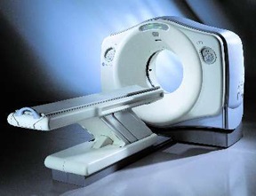
 CT (Computed Tomography) is a scan that produces a series of finely cut images capable of detecting a wide range of conditions that do not show or are not easily shown on conventional x-ray.
During the scan, a thin beam of x-rays is focused on the specific part of the body requested by your physician, such as the head, chest, liver, spleen, or spine. The x-ray tube is rotating rapidly around you as you lie on the scan table, but does not touch you. Using this rotation, multiple images are obtained as the beam passes through you at many different angles. This creates a cross-sectional image that is picked up by multiple detectors inside the scanner, recorded, and then relayed to a powerful computer. The computer then analyzes the information and constructs very detailed images of the body part in a variety of planes and formats (including 3D).
During certain CT scans, contrast medium may be given to enhance the study. One type is called barium sulfate, which is a thin, white mixture that you drink 30 – 45 minutes before certain abdominal scans. This is used to coat the intestine and allows greater visualization of the small bowel and large intestine. The other contrast medium is an iodine solution (commonly referred to as “dye”) that is given through an IV. This outlines the blood vessels and highlights various organs (e.g., liver, kidneys, spleen, etc.), so they may be more easily seen.
If this contrast is required, and you are 50 years of age or older, a certain blood test may be required to make certain your kidneys are functioning well enough to receive it.


